Rodríguez-Walker M et al., 2015, Biochem. J.
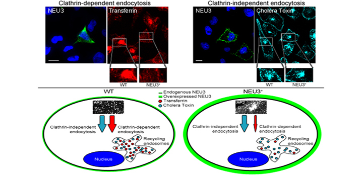
Gangliosides are sialic acid-containing glycosphingolipids mainly expressed at the outer leaflet of the plasma membrane. Sialidase NEU3 is a key enzyme in the catabolism of gangliosides with its up-regulation having been observed in human cancer cells. In the case of CME (clathrin-mediated endocytosis), although this has been widely studied, the role of NEU3 and gangliosides in this cellular process has not yet been established. In the present study, we found an increased internalization of Tf (transferrin), the archetypical cargo for CME, in cells expressing complex gangliosides with high levels of sialylation. The ectopic expression of NEU3 led to a drastic decrease in Tf endocytosis, suggesting the participation of gangliosides in this process. However, the reduction in Tf endocytosis caused by NEU3 was still observed in glycosphingolipid-depleted cells, indicating that NEU3 could operate in a way that is independent of its action on gangliosides. Additionally, internalization of α2-macroglobulin and low-density lipoprotein, other typical ligands in CME, was also decreased in NEU3-expressing cells. In contrast, internalization of cholera toxin β-subunit, which is endocytosed by both clathrin-dependent and clathrin-independent mechanisms, remained unaltered. Kinetic assays revealed that NEU3 caused a reduction in the sorting of endocytosed Tf to early and recycling endosomes, with the Tf binding at the cell surface being also reduced. NEU3-expressing cells showed an altered subcellular distribution of clathrin adaptor AP-2 (adaptor protein 2), but did not reveal any changes in the membrane distribution of clathrin, PtdIns(4,5)P2 or caveolin-1. Overall, these results suggest a specific and novel role of NEU3 in CME.
Authors: Rodríguez-Walker M, Vilcaes AA, Garbarino Pico-E, Daniotti JL.
Article: Rodríguez-Walker M et al., Biochem. J. (2015) 470, 131–144. doi: 10.1042/BJ20141550


